To understand the true nature of advanced endometriosis, one must look beyond the microscopic active implants and examine the structural havoc the disease wreaks inside the body. Endometriosis is fundamentally a chronic, progressive inflammatory disease. One of its most destructive consequences is the formation of pelvic adhesions—dense, fibrous bands of scar tissue that act like internal superglue, binding pelvic organs together. The surgical procedure to carefully cut and dismantle these painful bonds is known as Laparoscopic Adhesiolysis, or the release of adhesions.
How do these adhesions form? Every month, displaced endometriosis tissue bleeds inside the closed pelvic cavity. This internal bleeding triggers an intense healing response from the body’s immune system. Fibrin, a clotting protein, is deposited over the organs to contain the irritation. Over time, this fibrin matures into tough, vascularized collagen bands—scar tissue. In mild cases, these bands may simply link an ovary to the pelvic wall. In severe cases, they can lead to a condition known as a ‘frozen pelvis,’ where the uterus, ovaries, fallopian tubes, urinary bladder, and bowel are completely fused together into a single, immobile mass.
Pelvic adhesions are a major source of two primary symptoms: chronic pelvic pain and infertility. Anatomically, organs like the uterus and bowel are designed to slide smoothly past each other during movement, digestion, or sexual intercourse. When they are glued tightly together, any movement stretches and pulls on these highly sensitive tissues, triggering a sharp, tugging, or chronic aching pain. Adhesions also trap the ovaries and fallopian tubes, preventing them from interacting normally, which directly contributes to mechanical subfertility.
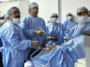
The laparoscopic release of adhesions requires exceptional anatomical knowledge and patience. The surgeon must carefully navigate a pelvis where normal anatomical landmarks have completely vanished. Using high-definition 4K magnification, we perform micro-dissection to separate the adhered organs layer by layer. This process must be carried out with extreme care, particularly when separating the bowel from the back of the uterus (the pouch of Douglas). We use ‘cold scissors’ (dissection without heat) and precision lasers to cut the scar tissue cleanly, preventing thermal damage to the underlying organs.
Once the organs are successfully untangled and restored to their normal, free-flowing positions, the surgeon’s focus shifts to preventing the scar tissue from returning. During the healing process, the body naturally wants to form new adhesions. To counteract this, we perform thorough pelvic irrigation with cold fluids to remove all blood clots and inflammatory factors. We then apply state-of-the-art anti-adhesion barriers—biocompatible gels or films that coat the raw surgical surfaces, acting as a physical shield for the first few critical days of healing. By freeing the pelvis from these restrictive internal bonds, laparoscopic adhesiolysis restores organ motility, delivers immense pain relief, and reopens the pathway for natural fertility.